Pour 12 ml of fresh, well-mixed urine into each URISED test tube. The amount of urine can be smaller in some cases (pediatric samples, some clinical conditions). Volume is to be measured and sample to be marked accordingly as the results are valid for this quantity.
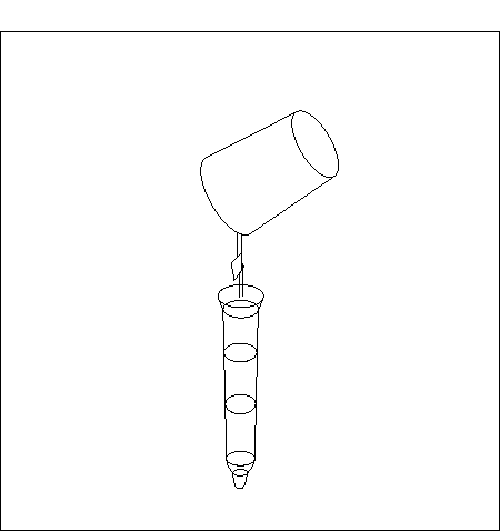
Close the test tubes with stoppers and expose them to centrifugation force.
Recommended time is 5 min at RCF 400×g. RCF for your centrifuge can be derived as:
RCF = 1,118 x 10-5 x r x RPM2, where RCF represents relative centrifugal field (g), r equals to radius (half-diameter) of the centrifuge, and RPM represents rotation speed in revolutions per minute.
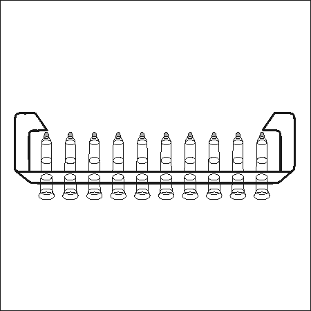
Remove the stoppers. Place the test tubes into the URISED stand and decant the supernatant with a single inversion.
Place the test tubes upside down on clean cellulose for 3 to 5 seconds.
Return the test tubes into upright position.
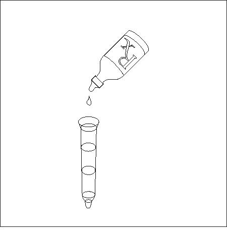
Add a single drop of URISED coloring substance to the sediment.
Mix the sample with the pipette tip attached to a corresponding pipette. The pipette tips are enclosed.
Use the same pipette to apply a drop of the sample to the semicircular opening on the URISED slide chamber.
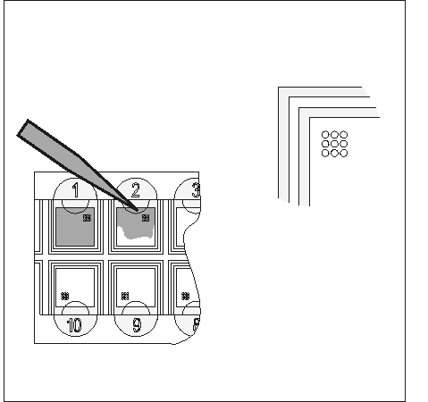
by means of capillary drawing the whole chamber becomes filled with the sample.
Circles located in each chamber assist evaluation of the sample. The volume of the sample within each circle is ensured by a fixed distance between both slides and equals 0,011 μl.
Cells in the sample are colored instantly; the colored sample is then stable approximately one hour at room temperature.
Cells in sediment are colored in the following way:
Examine the urine sediment with 100 x and 400 x objectives.
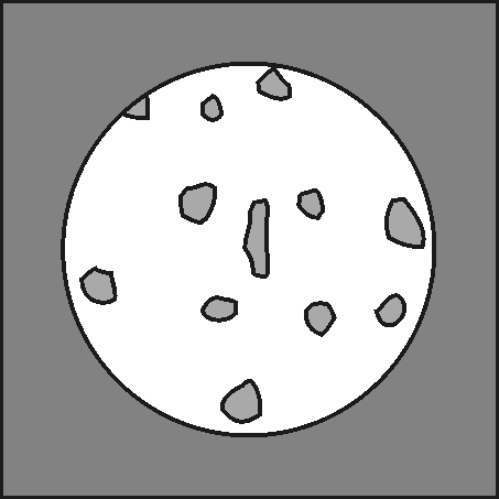
Evaluate the presence of cylinders at smaller magnification. Evaluate the presence and the number of cells at larger magnification. Circles may be used to assist the evaluation; each circle corresponds to a microscope field of view at 400× magnification.
Use an average number based on more measurements as a final result.
All the biological samples and used kit parts are to be handled according to the polices and procedures of your facility.
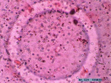
100×magnification: erythrocytes and leukocytes
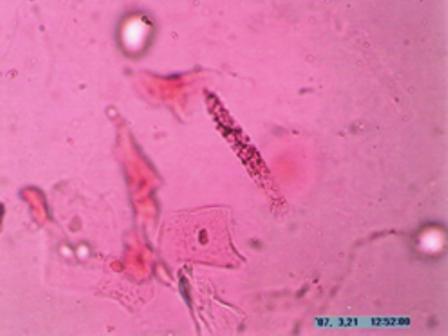
400×magnification: granulated cylinder (middle), hyaline cylinder (right above), epithelial cells of erythrocyte (bright, circular)
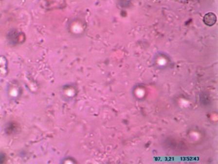
400×magnification: erythrocytes, bacteria, leukocyte (right above)
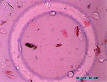
100×magnification: granulated cylinder (left)
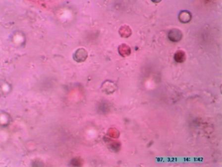
400×magnification: erythrocytes (bright) and leukocytes (dark), bacteria
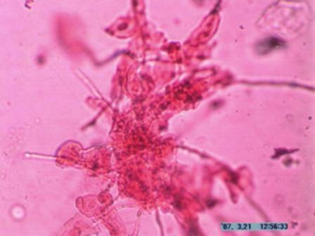
400×magnification: hyphae of fungus, erythrocyte (left below), epithelial cell (right above)
